
Understanding Electrocardiography – By Dr. Gnana Sankaralingam
 An electrocadiogram (ECG) is a recording of the electrical activity of the heart each time it contracts. Heart is a muscular organ whose purpose is to pump blood to tissues to supply them with oxygen. This is accomplished by four chambers, two smaller upper ones called Atria and two bigger lower ones Ventricles. Right atrium receives blood from the body while left atrium receives oxygenated blood from the lungs. They pump blood into right and left ventricles respectively. When ventricles contract, to prevent blood coming back to atria there are valves called mitral on left side and tricuspid on right side. Ventricles pump blood out of the heart, right to the lungs and left along entire circulatory system. To prevent blood returning back to ventricles there are valves, pulmonary on right side and aortic on left side. This is termed the cardiac cycle which outlines mechanical activity of the heart.
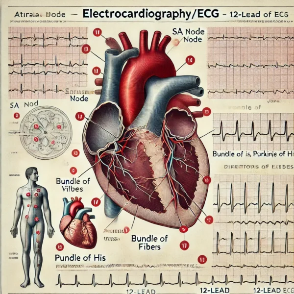
This mechanical function is initiated by an electrical conduction system, which contains wiring made of specialised tissues necessary to maintain rhythmic contraction. The system consists of Sinoatrial (SA) node, Atrioventricular (AV) node, pathways between them, bundle of His which divides into right and left bundle branches (left divides into anterior and posterior fascicles) both ending in Purkinje fibres in cardiac muscles. Impulses originate from SA node called natural pacemaker, which is under control of the brain through sympathetic and parasympathetic nerves in regulating rate of heart beat. Impulses spread through both atria by way of internodal pathways, causing both atria to contract and discharge electricity (depolarisation) simultaneously. Waves arrive at AV node located at right side of the septum between the atria, where it is delayed before reaching bundle of his. This phenomenon is to give time for ventricles to fill to capacity. From bundle of His whch is located just above the right ventricle, it travels to right and left bundle branches. Right one supplies right ventricle while left anterior supplies anterior and superior parts and left posterior supplies posterior and inferior parts of left ventricle. Impulses then activate purkinje fibres causing ventricles to contract and discharge electricity (depolarisation). Once blood is pumped out, ventricles relax to refill and get recharged (repolarisation) to continue the electrical cycle.
To record this, one needs electrodes placed on the body, and paper to get the tracing. 10 electrodes are placed on designated areas and with various combinations, twelve different views of electrical activity are demonstrated on ECG. Electrodes consist of limb and chest leads looking at the heart in frontal and horrizontal planes respectively. For limb leads, an electrode each is placed in left and right wrists and left ankle. One placed in right ankle is to stabilise the ECG, and does not take part in recording. Limb leads could be standard called bipolar as they have two electrodes, or be augmented as they comprise of three electrodes. In standard leads, one is positive and other negative and the difference in potential between them is recorded by ECG. In lead 1 right arm is denoted as negative and left arm as positive, in lead 2 right arm is made as negative and left leg as positive and in lead 3 left arm is made as negative and left leg as positive. L1 records electrical activity of the heart from lateral end while L2 and L3 record electrical activity of the heart from the inferior position.
Augmented leads are considered as unipolar as they comprise of one positive electrode recording potential at that point with reference to the other two. Because of this manner in which they are arranged, voltage is extremely low which must be augmented to equal the voltages of remainder of the ECG, which is accomplished by the machine. In lead AVR (Augmented Voltage of Right arm), right arm is the positive electrode in reference to left arm and left leg, recording electrical activity from the direction of right arm. In lead AVL (Augmented Voltage of Left arm), left arm is the positive electrode in reference to right arm and left leg, recording electrical activity from the direction of left arm. In AVF (Augmented Voltage of left Foot), left leg is the positive electrode in reference to left arm and right arm, recording electrical activity from the direction of bottom of the heart. There are precordial (chest) leads which are placed in front of the chest at the level of the heart. There are six of them usually, but if necessary tracing may be done with four more at the back.
Paper for recording consists of big squares, each divided into five small ones. Machine is run at a speed such that 300 big squares go through in one minute, which means that time taken by one big square is 0.2 secs and that by a small square is 0.04 secs, which is helpful in calculating the rate of heart beat as well as duration of the waves. Vertically each big square is 5mm in height and small one is 1mm in height which is useful in measurement of voltage. When recording, there is a base line (Iso-electric line) and deflection of the stylus above it are positive and below it are negative. Wave going toward an electrode records an upward deflection, wave going away from an electrode records a downward deflection and wave moving at right angle to an electrode will either cause a very small deflection or none. Each of the 12 leads views the heart from a different angle and as such each has a separate distinct pattern. Average direction and magnitude of electrical discharge (depolarisation) is denoted by mean vector, which for both atria and ventricles point downward and to the left toward lead 2. Thus lead 2 is used for monitoring patient in intensive care unit.
Once the ten electrodes are in proper places, they are attached to respective leads and connected to the machine. While patient remains still, recording is done for all 12 leads. To ensure that tracing is correct as to voltage, baseline is calibrated to deflect 10mm (2 big squares) with 1mv of current, which is termed standardisation. Interferences causing abnormal markings should be eliminated, which may be of AC interference from machines like ventilators, muscular tremors as in Parkinson’s or poor electrode contact with patient’s skin. ECG recording may be done while patient is resting, wearing portable machine for long period monitoring or as part of stress test. ECG is indicated in conditions like irregular heart beat, shortness of breath on exertion, suspected heart attack, valve problems and very high blood pressure. One must remember that ECG is a basic investigation, for it may not show everything and additional tests may be needed for complete assessment. Electrical activity is designated in the tracing as ‘P’ for atrial discharge, ‘QRS’ for ventricular discharge and ‘T’ for ventricular recharge, while atrial recharge which happens during ventricular discharge is hidden. Tracing is done for few P-QRS-T cycles in each of the 12 leads to complete the ECG. Recording is looked through and interpreted as to whether it is normal or not.